
Figure 2.
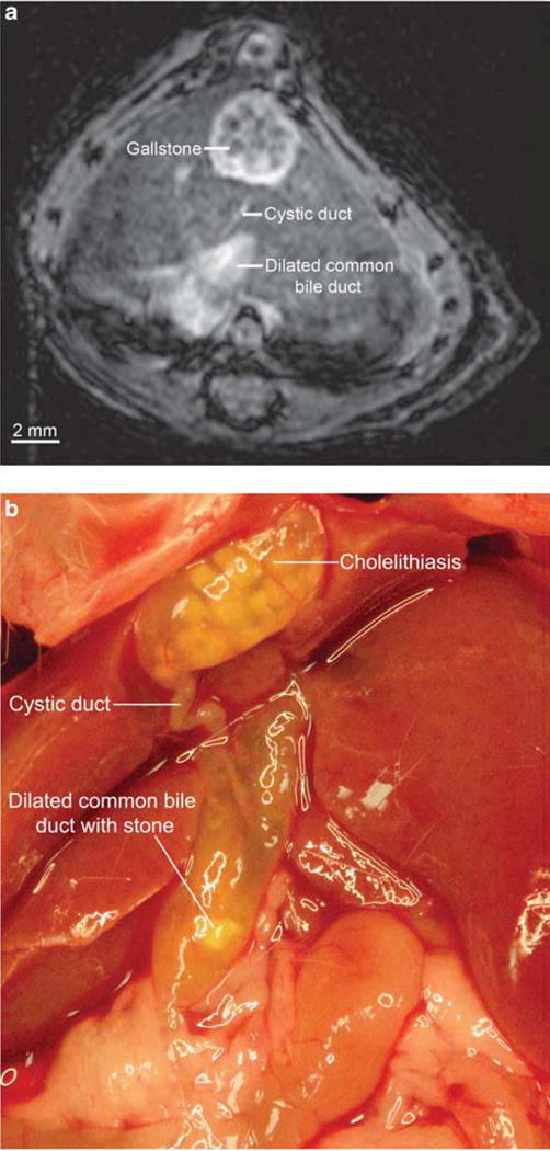
Cholelithiasis and dilated common bile duct in mdr2 knockout (mdr2KO) mouse seen with magnetic resonance (MR) imaging and confirmed by anatomic dissection. (a) Gallstones are seen as 0.35 mm, circular hypointensities. Common bile duct is dilated to over 1 mm, whereas the cystic duct appears to be of normal or slightly enlarged caliber (IntraGateFLASH (IntraGate Fast Low Angle Shot), echo time 3 ms, repetition time 82 ms, 30 repetitions, image matrix 256 × 256 pixels, acquisition matrix size 128 × 128 pixels, resolution 125 × 125 μm, slice thickness 250 μm, acquisition time 5.3 min). (b) Dissection of the same mouse immediately after imaging demonstrates extensive cholelithiasis and a severely dilated common bile duct with choledocholithiasis, consistent with MR findings.