
Figure 3.
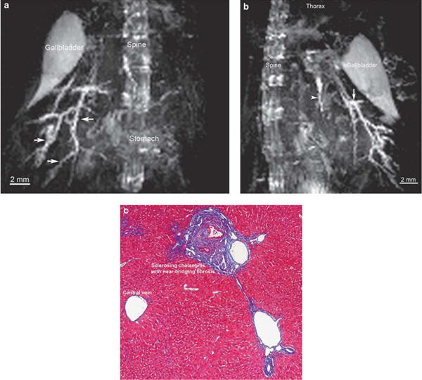
Magnetic resonance (MR) imaging with maximum intensity projection (MIP) post-processing demonstrates intrahepatic changes consistent with sclerosing cholangitis in mdr2 knockout (mdr2KO) mouse, as corroborated by liver histology. (a) Coronal view of mdr2KO mouse biliary tree using MIP algorithm demonstrates dilated, diffusely irregular intrahepatic ducts (arrows) (TurboRARE (Turbo Rapid Acquisition with Relaxation Enhancement) with respiratory triggering, echo time 8 ms, effective echo time 24, RARE factor 8, image and acquisition matrix 256 × 128 × 128 pixels, resolution 200 μm isotropic, acquisition time 39min with respiratory rate of ∼70breath/min). (b) On sagittal view, the same MIP demonstrates additional features, including beading (arrow) and a sclerotic left ductal system, hallmarks of primary sclerosing cholangitis. (c) Trichrome-stained section of mdr2KO mouse liver (× 10) demonstrates characteristic features of sclerosing cholangitis, consistent with MR findings.