
Fig. 2. NoV infection requires RNAi suppression.
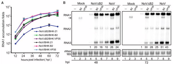
(A) BHK-21 cells or BHK cells stably expressing B2 or VP35 were mock-infected or infected by NoVΔB2 or NoV of the same titer. Every 12 hours postinfection (hpi), the viral genomic RNA1 levels were determined by quantitative RT-PCR with the accumulation level of NoVΔB2 in BHK-21 cells at 12 hpi set as 1. Error bars indicate standard deviation of three replicates. (B) Accumulation of NoV and NoVΔB2 RNAs 1 to 3 in the infected cells detected by Northern blotting. RNA1 signal quantified by phosphorimaging was shown with that of NoVΔB2 in BHK-21 cells (lanes 4) set as 1. Detection of B2 transgene mRNA (arrow) was visible. 18S rRNA staining served as loading control.