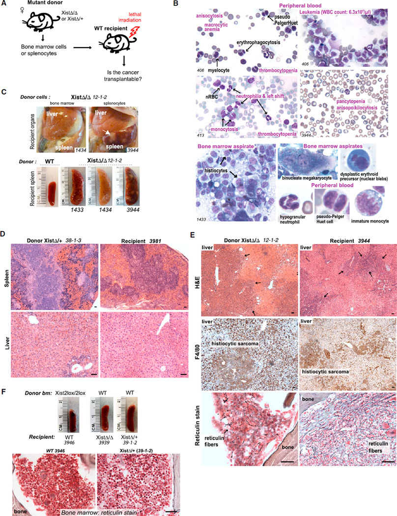
Figure 5. Transplantability of MPN/MDS Suggests a Hematopoietic Rather Than Stromal Origin.
(A) Schematic of transplantation experiments.
(B) Dysplasia in peripheral blood and bone marrow of mutant-to-WT transplants. See also Figures S3A–S3C.
(C) Histiocytic sarcoma recapitulated in WT recipients of mutant bone marrow or splenocytes. Recipient livers and spleen (1434) were pale from anemia. White masses in recipient spleen contain histiocytic sarcoma (arrow). Mice succumbed within 40 days after transplantation.
(D) H&E stain of EMH in matched donor and recipient livers and spleens, as indicated.
(E) Immunohistochemistry confirms histiocytic sarcoma in donor and recipient (middle panels). Reticulin stain of bone marrow shows exuberant myelofibrosis in donor (at necropsy) and recipient (40 days after transplantation).
(F) WT-to-mutant transplantations reversed disease. WT-to-WT transplantation controls were normal. Scale bars represent 100 µm. See also Figure S4.

