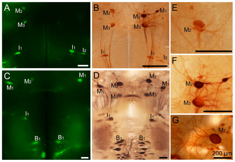
Figure 2. Double-labeling by FLICA and neurofilament immunohistochemistry in whole-mounted lamprey brains at 2 weeks post spinal cord transection.
A, FLICA labeling in the whole-mounted brain from a larval lamprey (12 cm, approximately 4 year old). FLICA-labeled identified neurons have been marked. B, The immunohistochemical staining of neurofilaments performed after FLICA staining (the same animal in A). The morphology of identified neurons has been kept intact and the distribution of neurofilaments is normal. C, FLICA staining in the whole-mounted brain from a post-metamorphic adult lamprey. D, The immunohistochemical staining of neurofilaments conducted after FLICA labeling (the same animal used in C). The distribution pattern of neurofilaments is normal. E, F, Higher magnification images of identified neurons from B. G, Higher magnification images of identified neurons M2 from D. The identified neurons have been kept intact in both lamprey brains. Scale bar: 200 μm.

