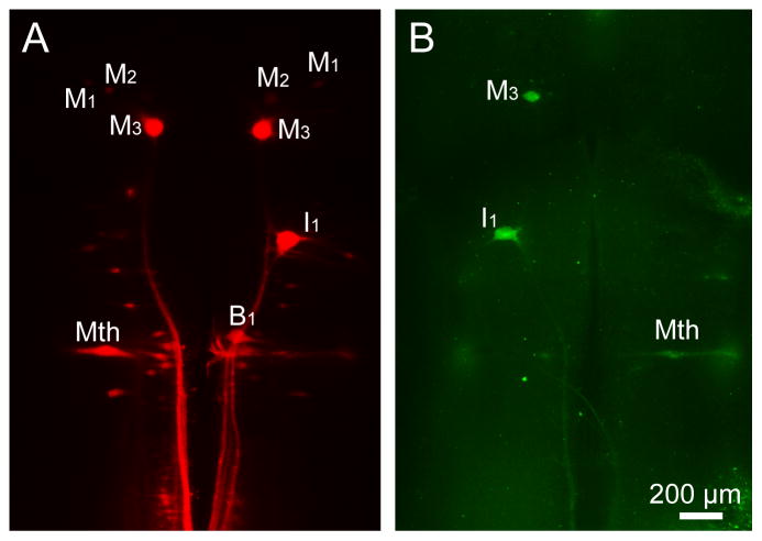
Figure 4. Double-labeling by FLICA and retrograde labeling tracer DTMR in whole-mounted larval lamprey brain at 2 weeks after spinal cord transection.
A, Retrograde labeling tracer DTMR (red) shows neurons in larval lamprey brain 2 weeks after application at the time of transection. B, Activated caspases in identified reticulospinal neurons, such as M1 and I1 neurons, revealed by FAM-VAD-FMK labeling (green, FLICA labeling) in the same brain used in A. Scale bar: 200 μm.

