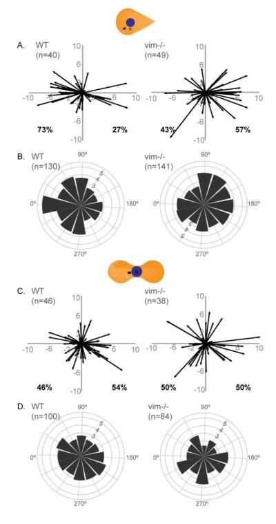
Figure 4. Absence of VIF affects cell polarity.
WT and vim-/- MEFs were seeded on 1000 μm2 micropatterns of teardrop (A, B) or dumbbell (C, D) shapes. The cells were stained with gamma-tubulin to mark the centrosome and Hoechst to locate the nucleus. The (x,y) coordinates for the center of the nucleus and center of the centrosome were calculated using ImageJ. The nucleus center – centrosome center vectors was then plotted for the teardrop (A) and dumbbell (C) shapes (See Materials and Methods). For each cell type/shape (B & D), the vectors were converted into angles and the number of samples in a given 30-degree increment was determined and the square root of this number was plotted on a polar coordinate plot (See Materials and Methods). n=number of cells used to generate the plots.

