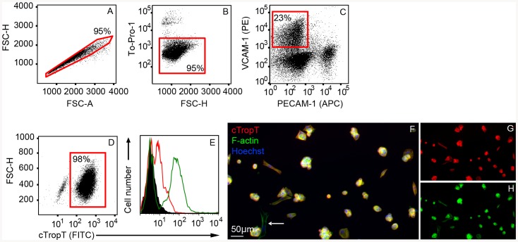
Figure 2. Purification of embryonic cardiomyocytes by Flow Cytometry.
Flow Cytometry plots of E10.5–E11.5 cardiac cells labeled with specific antibodies to VCAM-1 and PECAM-1 (A–C). Cells are gated and sorted based on doublet discrimination (A), viability (B) and VCAM-1 positive PECAM-1 negative population (C). Flow Cytometry plot of sorted fixed cells, stained with cTropT antibodies to verify cardiac identity (D). Flow Cytometry histograms (overlays) showing control cTropT staining of neonatal hearts (E). Un-stained cells (black), isotype control (red outline) and neonatal heart cells (green outline). The percentage of gated cells through each step of the sort is indicated in each plot. Immunofluorescence staining (cTropT) of sorted cells cultured on gelatin coated slides for two days to verify cardiac identity and cell viability (F). F-actin and cTropT in separate channels (G–H). A low frequent cTropT-negative cell is indicated (arrow).

