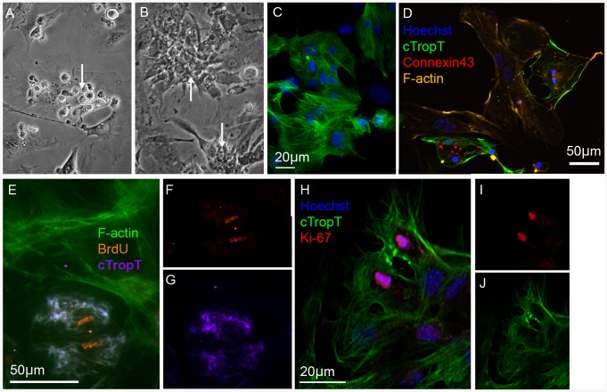
Figure 6. In vitro culture of primary embryonic cardiomyocytes after FACS.
Phase contrast images of FACS-isolated cells grown on irradiated embryonic cardiac fibroblasts for two (A) or six (B) days. Rounded cells attached to the fibroblasts (A) and beating clusters of cardiomyocytes (B, C). Immunofluorescence images of the co-cultured cells (C–J). The FACS-isolated cells form a tight meshwork of beating cardiomyocytes in close contact with the surrounding fibroblasts as well as expressing cTropT (C) and Connexin43 (D). Co-cultures labeled with BrdU after five days for 24 h (E–G). Incorporation of BrdU is detected in cardiomyocytes but not in the surrounding irradiated fibroblasts. A optical section of a dividing cardiomyocyte in co-culture with fibroblasts for five days visualized by Ki-67 expression (H–J).

