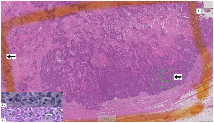
Figure 1. Snapshot of WSI showing the annotated areas for scoring mitosis microscopically and digitally.
1- An overview image of the WSI, 2- The defined area for counting mitosis microscopically, 3- The defined area for counting mitosis digitally, 4a- Microscopic snapshots of different mitotic figures, 4b- Digital snapshots of different mitotic figures.

