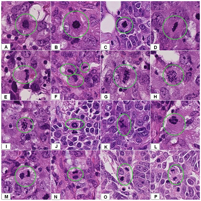
Figure 2. Snapshots from several WSI of several breast resections diagnosed previously as an infiltrative ductal carcinoma using a light microscopy.
These snapshots showing different appearances of mitotic figures encircled by green circles. Panels A–C show cells in early metaphase. Panels D–G show different forms of mitotic division in late metaphase. Panel H–L shows different forms of anaphase. Panels M–P show cells in telophase.

