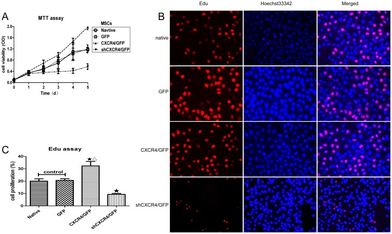
Figure 3. Regulation of CXCR4 expression affects the proliferation of MSCs in vitro.
(A) Viability was decreased in MSCsshCXCR4/GFP and increased in MSCsCXCR4 /GFP compared to MSCsGFP or MSCsnative (control), as measured by the MTT assay (Mean ± SD; n = 6; P<0.05 from NO. 4 to 5 time point). (B) All cell nuclei exhibited blue fluorescent Hoechst 33342 staining, and EdU labeling indicated replicating cells. In MSCsCXCR4, an increased number of cells exhibited red fluorescent EdU labeling following CXCR4 overexpression. In MSCsshCXCR4/GFP, fewer cells exhibited red fluorescence, indicating that EdU labeling decreased with CXCR4 knockdown. (C) The percentages of red fluorescent cells for different MSC groups are indicated in the histogram. There were more replicating cells for MSCsCXCR4 /GFP than the control group. ★P<0.05 vs. control, and ▵P<0.01 vs. MSCsshCXCR4/GFP.

