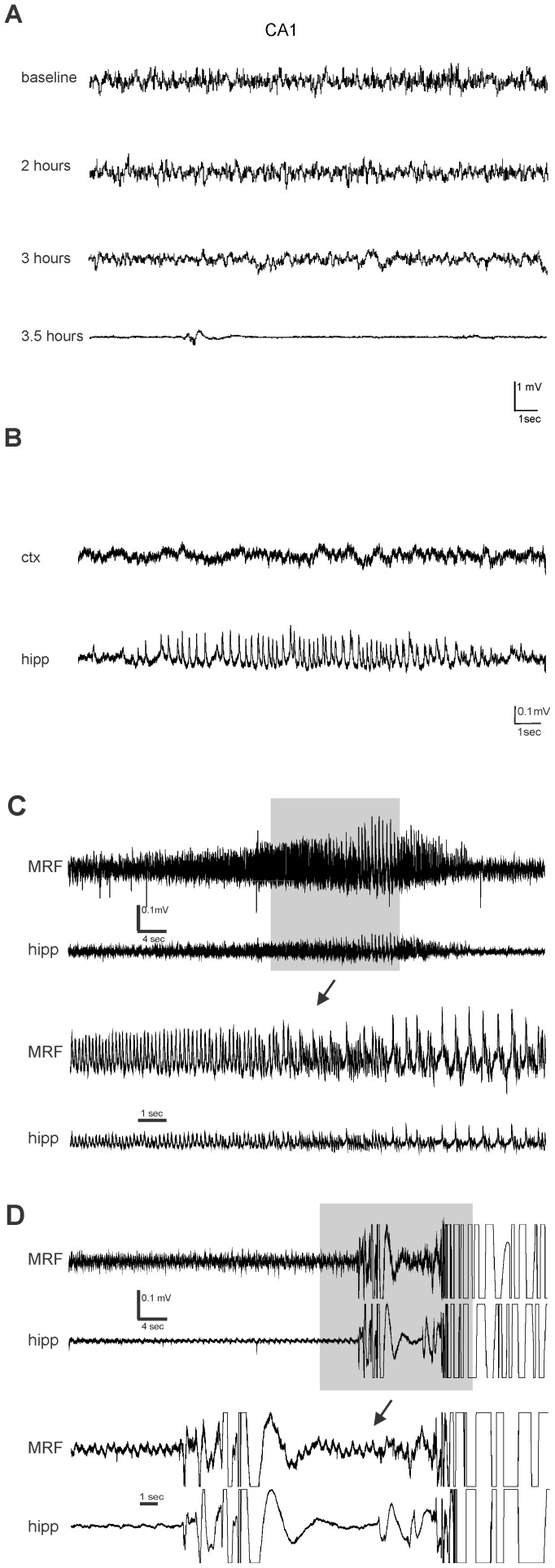
Figure 7. EEG abnormalities during hypoglycemia is not associated with seizure behavior.
A: Representative EEG recording of hippocampus (CA1) at baseline (post-fasting; prior to insulin IP), 2 hours, 3 hours (slower waves) and 3.5 (EEG suppression) hours after insulin IP B: Electrographic seizure activity observed in CA1 (lower trace) after suppression of EEG activity. No ictal activity in cortex (upper trace) C: EEG recording of hippocampus and contralateral MRF obtained in STZ rat during hypoglycemia. Electrographic seizure activity observed after suppression of EEG activity. Lower trace illustrates magnification of the area in the gray box D: EEG of the rat in (C) during a behavioural seizure; ictal activity may be masked by movement artifact.

