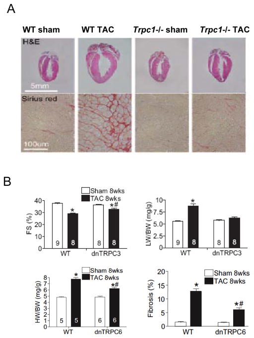
Figure 1.
A) Effect of gene deletion of TRPC1 on in vivo mouse heart subjected to pressure overload. Panels show baseline and post aortic constriction hearts (top) and myocardial histology (lower) for both control and TRPC1−/− hearts. There was less hypertrophy, chamber enlargement, and fibrosis in hearts lacking TRPC1. B) Mice with myocyte-selective expression of dominant negative TRPC3 display less decline fractional shortening (%FS) and less hypertrophy (LV/body weight, LW/BW) with sustained pressure overload (TAC). Mice with myocyte-targeted overexpression of a dominant negative TRPC6 display less cardiac hypertrophy in response to TAC. There is also less interstitial fibrosis. From Wu et al [48] with permission. C) Mice with cardiac myocyte targeted TRPC3 overexpression develop dilated and depressed heart function after 8 months, with enhanced stimulation of NFAT. From Nakayama et al [46] with permission. D) Myocytes subjected to transverse aortic constriction (TAC) display enhanced store-operated calcium entry, whereas this is blocked in 90% of cells studied from mice E).

