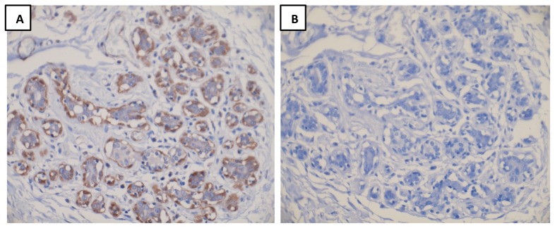
Figure 1. Dicer expression by IHC in normal breast tissue.

Representative images of Dicer staining (A) without blocking peptide and (B) with blocking peptide are shown. Panel A is representative of Dicer staining in normal breast tissue.

Representative images of Dicer staining (A) without blocking peptide and (B) with blocking peptide are shown. Panel A is representative of Dicer staining in normal breast tissue.