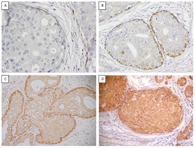
Figure 2. Dicer expression in DCIS.

Representative images of the spectrum of the staining intensity observed for Dicer in DCIS where 0 = negative (A), 1 = weak (B), 2 = moderate (C) and 3 = strong (D).

Representative images of the spectrum of the staining intensity observed for Dicer in DCIS where 0 = negative (A), 1 = weak (B), 2 = moderate (C) and 3 = strong (D).