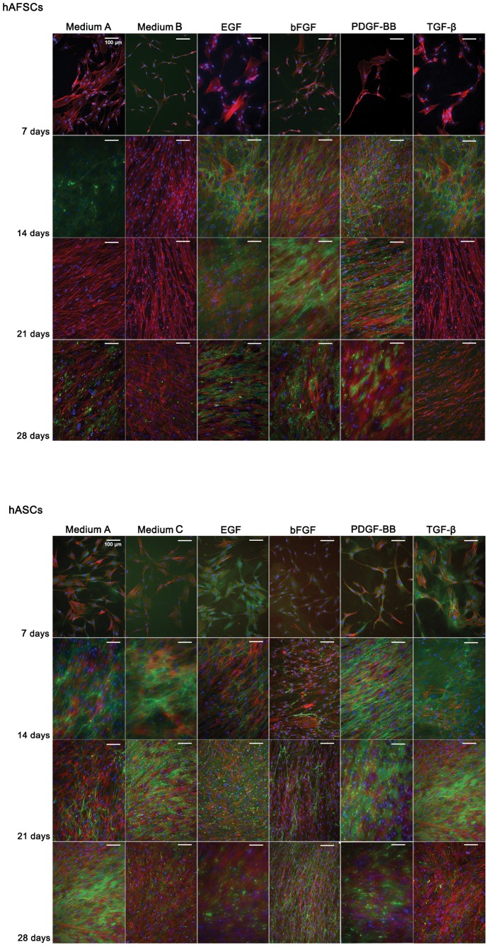
Figure 2. Tenascin C immunolocation in hAFSCs and hASCs cultured up to 28 days in different supplemented media.
DAPI (blue)and phalloidin-conjugate (red) stain cell nucleus and cytoskeleton, respectively. Tenascin C is stained in green and represents a tendon ECM protein. Scale bar represents 100 µm. Magnification: 200 x.

