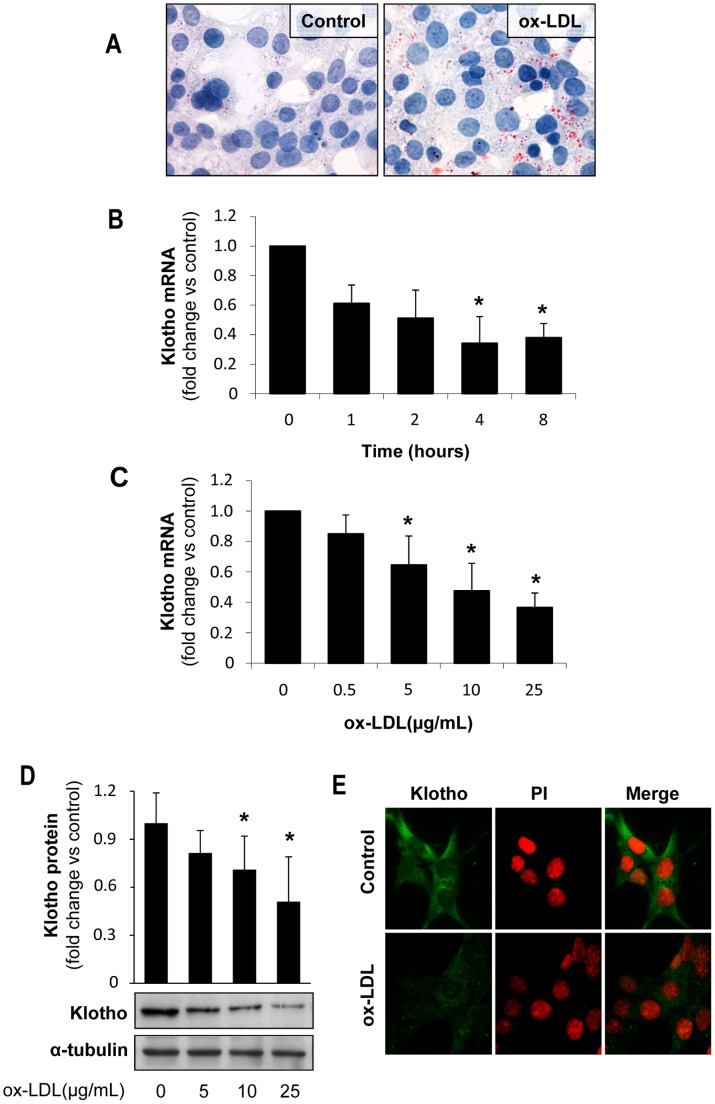
Figure 4. Oxidized LDL decrease Klotho expression in cultured tubular cells.
(A) Oil-Red-O staining in murine proximal tubular cells (MCT) showing increased lipid accumulation after 24 h incubation with ox-LDL (25 µg/mL). Ox-LDL decreases Klotho mRNA expression, as determined by quantitative RT-PCR, in a time (B) and dose-dependent manner (C) in proximal tubular cells (MCT). Mean±SD of three independent experiments. *p<0.05 vs control. Klotho protein expression, as determined by Western blot (D) and confocal microscopy (E), in MCT treated with ox-LDL (25 µg/mL) for 24 h. Indirect immunofluorescence using anti-Klotho with secondary Alexa Fluor 488–conjugated antibody (green). Nuclei were stained with propidium iodide (PI, red). Images are representative of three independent experiments.

