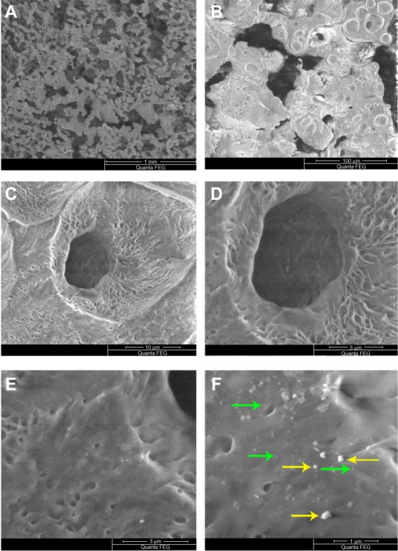
Figure 1.

Scanning electron microscopy images of PCL-RNP. (A) PCL-RNP showing pores in the range of 50–100 μm. (B–D) Further magnification into the scaffold. (E and F) Nanoparticles and agglomerates embedded on the surface of the scaffold.
Note: Green and yellow arrows show embedded nanoparticles and agglomerations, respectively.
Abbreviations: PCL, polycaprolactone; RNP, resveratrol-loaded albumin nanoparticles.
