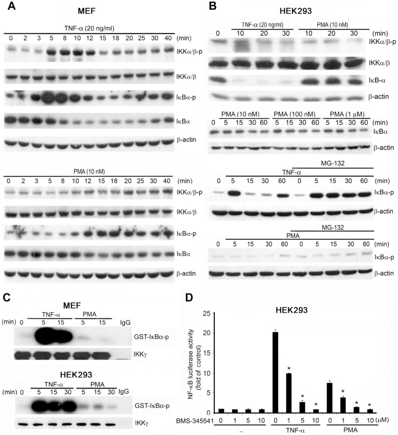
Figure 3. PMA induces weaker IKK and NF-κB activation than does TNF-α.
MEFs (A) and HEK293 cells (B) were treated with TNF-α (20 ng/mL) or PMA (10 nM) for the indicated periods. In some experiments (B), HEK293 cells were pretreated with MG-132 (10 µM) for 30 min prior to treatment with PMA at the concentration indicated or with TNF-α (20 ng/mL) for various periods. Total cell lysates were collected and subjected to western blotting analysis; the antibodies used are indicated. The western blots are representative of 3 independent experiments. (C) After treatment with TNF-α (20 ng/mL) or PMA (10 nM) for the indicated times, IKK complex activity was determined using an in vitro kinase assay in which GST-IκB was included as the substrate. The autoradiograph shown here is representative of 3 independent experiments. (D) HEK293 cells transfected with the κB reporter andβ-galactosidase plasmids were treated with the IKK inhibitor BMS-345541 (1–10 µM) for 30 min and then stimulated with TNF-α (20 ng/mL) or PMA (10 nM) for 8 h. NF-κB luciferase activity was determined and normalized relative toβ-galactosidase activity. Data are the means ± SEM from 3 independent experiments. * P<0.05 indicates significant inhibition of TNF-α and PMA responses after treatment with BMS-345541.

