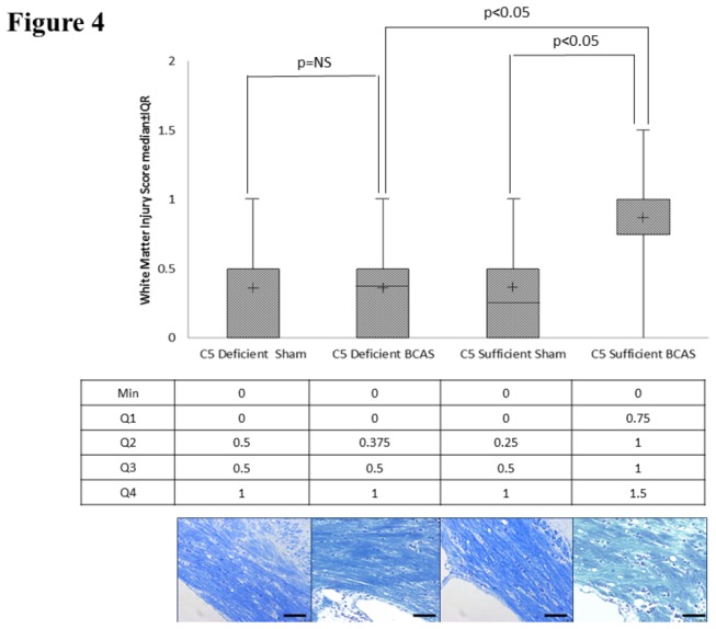
Figure 4. White matter ischemia.

Kluver-Barrera staining for white matter ischemic changes in the medial corpus callosum in C5 sufficient and C5 deficient mice subjected to sham and BCAS operations. Above: White matter injury scores. Middle line is the median. The (+) is the mean. The upper and lower lines on the box are the 75% and 25%, respectively. The uppermost and lowermost bars are minimum and maximum, respectively. Values in the text are expressed as median and interquartile range Below: Representative coronal sections of the right corpus callosum with high magnification insert (medial corpus callosum). Bars indicate 50µm. n=10 C5D/ BCAS, n=10 C5D/ sham, n=9 C5S/ BCAS, n=9 C5S/ sham.
