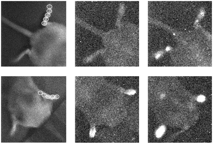
Figure 2. Image of left and right hind paws (upper panel) and front paws (lower panel) of experimental mouse 7.
In both rows, the left image is a position image, recorded under weak light illumination before the actual imaging of UPE. Middle images represent UPE before luminol injection. Right images represent UPE immediately after luminol injection.

