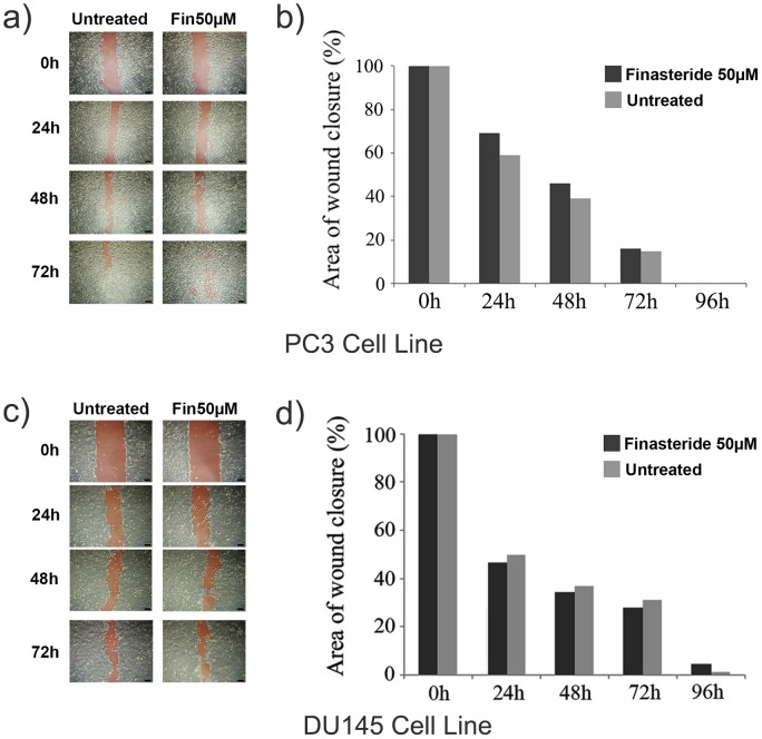
Figure 5. Finasteride’s effects on the migration of the human prostate cell lines PC3 and DU145 in the wound closure migration assay.
Cells were cultivated in 6-well plates at 4×10⁴ cells/cm2 until 100% confluence was achieved. The monolayers were scratched in a straight line, and the wounded monolayers were then given culture medium with or without 50 µM finasteride. The wound area was inspected after 24, 48, 72 and 96 hours using an inverted phase contrast microscope with a digital camera. a) Representative images showing the wound closure of finasteride-treated and control PC3 cells at 0 to 72 hours. Red highlighted areas represent the open-wound area. Scale bar = 50 µm. b) The initial red highlighted areas were measured using the ImageJ™ software, and the remaining areas were calculated as a percentage of the initial wound area. A wound-closure graphic was made by dividing the area values that were obtained at the indicated time points for the control and finasteride-treated cells. Finasteride slightly inhibited cell migration; however, this difference was not statistically significant (p>0.05). c) Representative images showing the wound closure of finasteride-treated and control DU145 cells at 0 to 72 hours. Red highlighted areas represent the open wound area. Scale bar = 50 µm. d) As observed for the PC3 cells, finasteride slightly inhibited cell migration (p>0.05).

