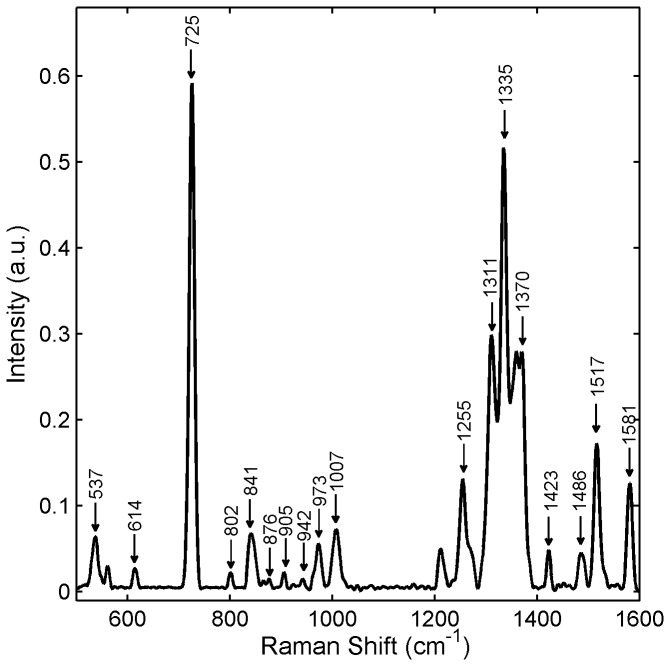
Figure 2. Raman spectrum of 1%w/w Tenofovir in water.
1% Tenofovir solution was prepared in alkalinized water (50 mM NaOH). The Raman spectrum of the Tenofovir solution was acquired using a Horiba LabRam ARAMIS Raman microscope with an excitation wavelength of 633 nm, 100μm slit width and a 10x objective lens. The acquisition time was 60s with 3 accumulations. Data were normalized to the area under the curve.

