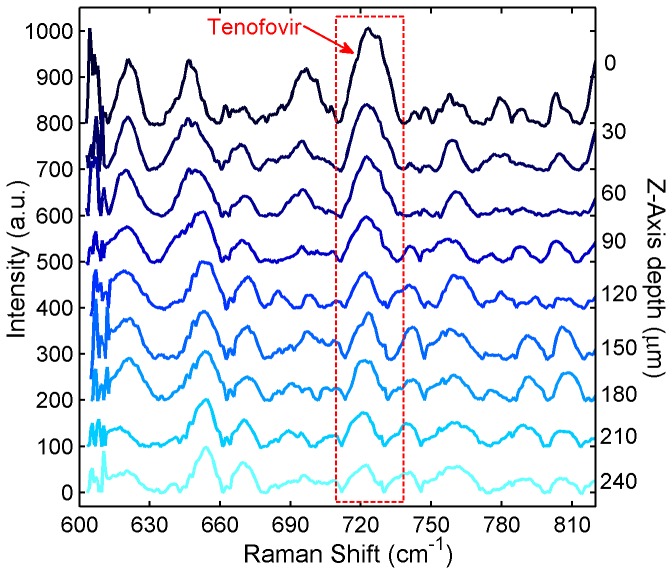
Figure 10. Z-scanning Raman spectra for Tenofovir penetrating excised tissue in the Transwell assay.
7 porcine buccal tissue specimens were incubated under an isotonic solution of 1% Tenofovir in PBS for 6 h in the Transwell assay. After incubation, the tissue was stored at −80°C overnight to stop the drug permeation process. Thawed tissue specimens were subjected to confocal Raman scans at different depths beneath the tissue surface to quantify Tenofovir distribution within the tissue specimens. The Raman spectra were acquired at different z-axis depths using a Horiba Xplora confocal Raman microscope with an excitation wavelength of 785 nm and an Olympus 50x long-working distance objective lens (N.A. =0.5). The acquisition time was 240 s for each spectrum. All spectra were normalized with respect to the tissue Raman band at 643 cm-1 and offset vertically for clarity of presentation.

