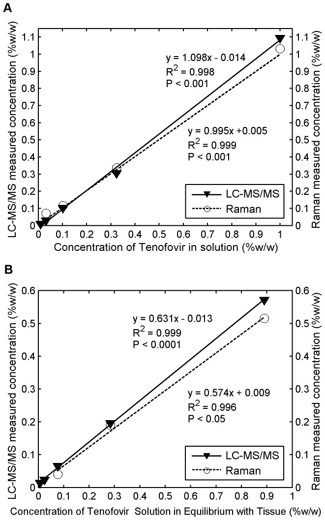
Figure 12. Comparative measurement of Tenofovir concentration using Raman vs.
LC-MS/MS in solution and tissue.
Freshly excised porcine buccal tissue specimens were incubated in serially diluted concentrations of Tenofovir in Ringer's solution and allowed to equilibrate for 24 h. After incubation, the tissue specimens and the surrounding fluid were collected and stored at −80°C overnight for analysis by both techniques: (a) The initial serially-diluted concentrations of Tenofovir in Ringer's solution measured by both techniques were plotted against its known concentrations in solution. These two techniques produced slopes that did not differ significantly (ANCOVA, P > 0.05); (b) the final concentrations of Tenofovir within tissue specimens measured by both techniques were plotted against concentrations of Tenofovir solution in equilibrium with tissue. ANCOVA also showed that the slopes obtained by both techniques were not significantly different (P > 0.05).

