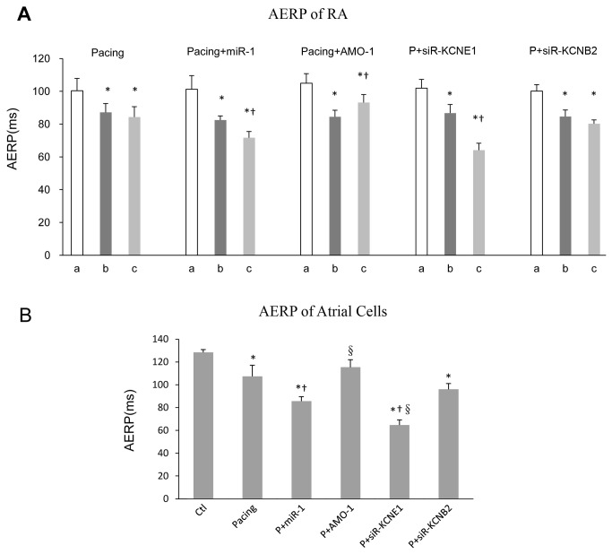
Figure 3. AERP evaluations.
AERP of RA obtained from animals as described in the Materials and Methods section (A). The AERP values (y axis: ms) were obtained at 3 time points: before pacing (“a”), before infection control/recombinant LVs (“b”), and 1-week after infection (“c”). * P < 0.05 vs. “a”; † P < 0.05 vs. “b”, one-way ANOVA and Tukey’s post-hoc test; n = 6 independent rabbits for each group. AERP of atrial cells were measured by whole cell patch clamp in vitro (B).* P < 0.05 vs. Ctl, † P < 0.05 vs. Pacing, §P < 0.05 vs. P+miR-1, one-way ANOVA and Tukey’s post-hoc test; n = 6 independent RNA samples for each group.

