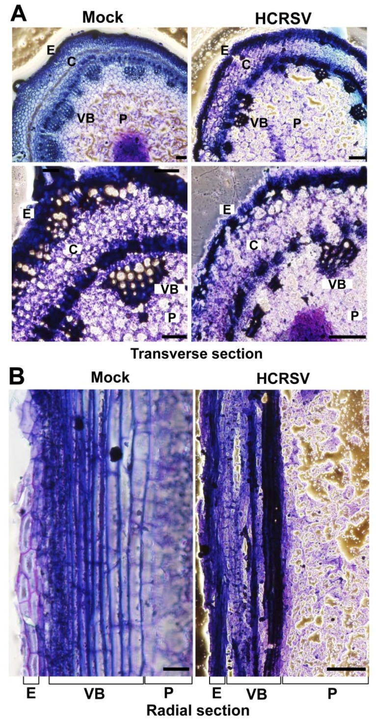
Figure 2. Comparison of transverse and radial sections of mock and HCRSV- infected kenaf stem at 15 dpi.

(A) Transverse section of mock and HCRSV-infected kenaf stems. The pith is located in the middle of the transverse section, surrounded by vascular bundles. (B) Partial radial sections of mock and HCRSV-infected kenaf stems. E, epidermis; VB, vascular bundle; C, cortex; P, pith. Bar = 100 µm.
