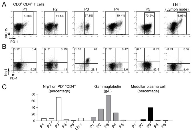
Figure 7. Nrp1 expression in malignant Tfh-like cells in AITL.
Cell suspensions from malignant lymph node biopsies of 5 AITL patients (P1-P5) and one non-malignant reactive lymph node (LN 1) were analyzed for CXCR5 and PD-1 expression (A), and for Nrp1 and PD-1 expression (B) in gated CD3+ CD4+ T cells. (C) Bar graph display of percentage of Nrp1+ cells among PD1+ CD4+ T cells, serum gammaglobulin concentration and medullar plasmacytosis (percentage plasma cells among bone marrow cells) for each patient. Note that only P3 shows a significant proportion of Nrp1+ malignant T cells, high gammaglobulinaemia, and important medullar plasmacytosis.

