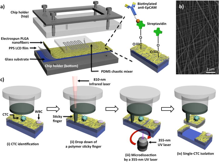
Figure 1.
a) The configuration of PLGA nanofiber (PN)-NanoVelcro Chip for the high-purity isolation of circulating tumor cells (CTCs) from prostate patients' peripheral blood samples. A custom-designed chip holder is employed to sandwich a PN-NanoVelcro Chip that is composed of (i) an overlaid PDMS chaotic mixer chip and (ii) a transparent PN-NanoVelcro substrate, prepared by depositing electrospun PLGA nanofibers onto a commercial laser microdissection (LMD) glass slide (with a pre-deposited 1.2-μm-thick PPS membrane). Streptavidin is covalently attached onto the PLGA nanofibers for conjugation of the capture agent (i.e., biotinylated anti-EpCAM). b) A SEM image of the electrospun PLGA nanofibers. c) Schematic illustration of the workflow for isolation of high-purity CTCs using a 4-step laser capture microdissection (LCM) technique: (i) CTCs (CK+/CD45-) are first identified from surrounding WBCs (CK-/CD45+), and an LCM cap (with a polymer membrane on bottom) is then aligned over the substrate, (ii) an IR-laser is used to melt the polymer membrane, dropping down a “sticky finger” for adhering onto the PN-NanoVelcro substrate, (iii) a 355-nm UV laser is utilized to dissect the substrates underneath the identified CTC, and (iv) by removing the LCM cap high-purity CTCs can be obtained for subsequent molecular analysis.

