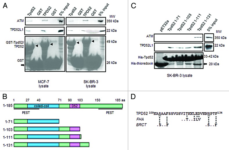
Figure 7. Interactions between TPD52 and ATM. (A) GST pull-down assays using GST-tagged full-length mouse Tpd52 or human TPD52 or GST tag, and MCF-7 (left) or SK-BR-3 (right) cell lysates. Upper and middle panels show the results of western blot analyses using ATM antibody and TPD52L1 antisera, respectively. The lower panel shows Ponceau S staining to reveal GST-Tpd52/TPD52 (arrowheads) or GST tag (arrow). At least 3 independent experiments were performed. (B) Schematic representations of deleted Tpd52 recombinant proteins. Amino acid coordinates for each motif/domain are indicated. (C) Pull-down assays using thioredoxin-6His-tagged Tpd52-deleted proteins and SK-BR-3 cell lysates. Upper and middle panels show results of western blot analyses using ATM antibody and TPD52L1 antisera, respectively. The lower panel shows Ponceau S staining to reveal His-Tpd52 proteins or His-thioredoxin tag (arrow). Results shown are representative of those obtained from 3 independent experiments. (D) Sequence of TPD52 aa 109–134 with broken lines indicating FHA and BRCT motifs as predicted through ELM. S111 and S131 are underlined.

An official website of the United States government
Here's how you know
Official websites use .gov
A
.gov website belongs to an official
government organization in the United States.
Secure .gov websites use HTTPS
A lock (
) or https:// means you've safely
connected to the .gov website. Share sensitive
information only on official, secure websites.
