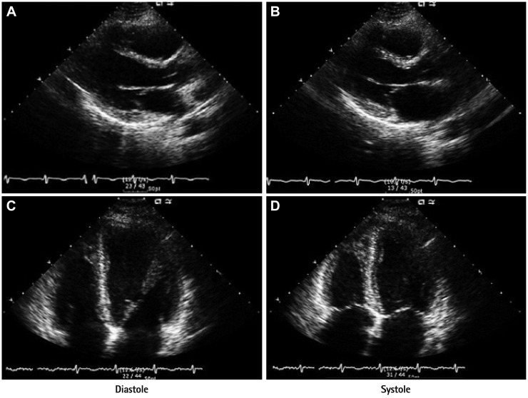
Fig. 3.
Transthoracic echocardiography at the time of pulmonary embolism shows severe left ventricular systolic dysfunction with hypokinesia of the base and mid ventricular segment and hypercontractility of the apex. A and B: parasternal long-axis view in diastole and systole. C and D: apical four-chamber view in diastole and systole.

