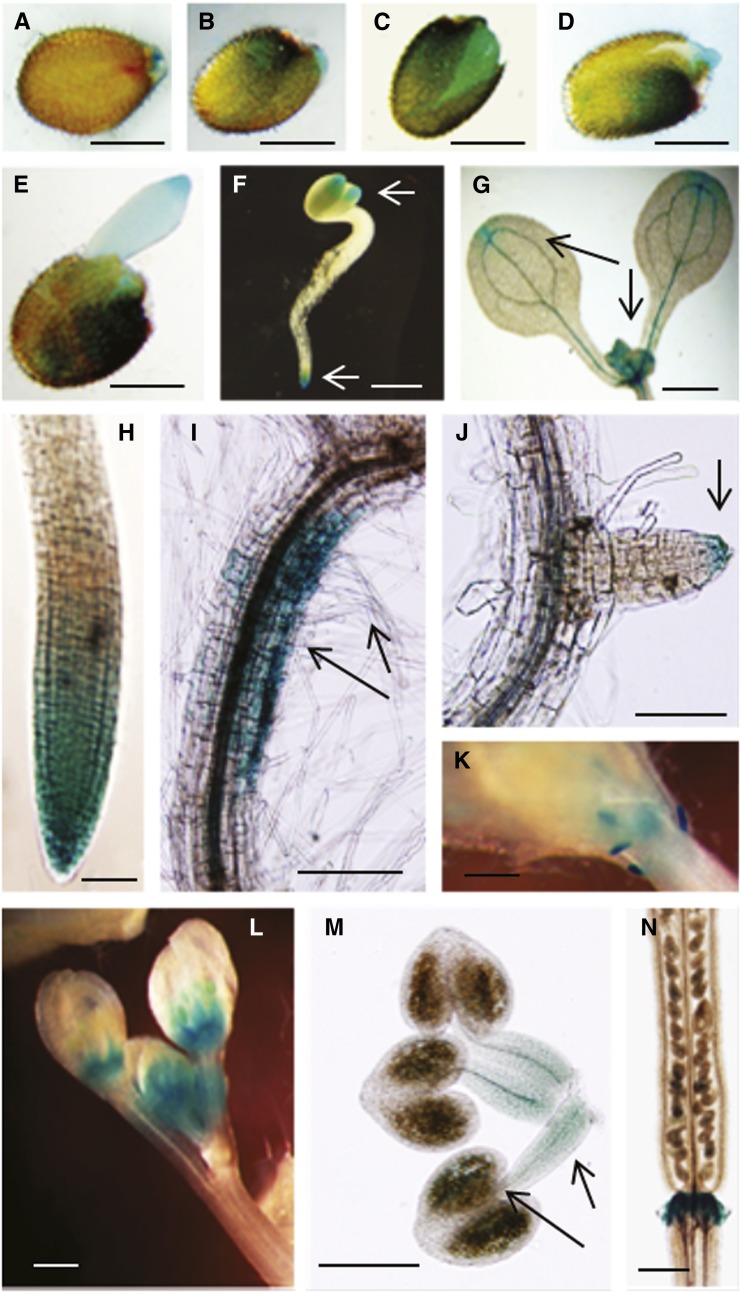
Figure 1.
Histochemical GUS Staining Showing Spatial and Tissue-Specific Expression Driven by the Arabidopsis CaM KMT Promoter.
(A) to (E) GUS expression in Arabidopsis (T2 generation seed harboring CaM KMTPro:GUS construct) seed at various times after imbibition after stratification. After surface sterilization, seed was stratified at 4°C for 2 d. GUS staining of seed immediately after stratification (A), 1 d after stratification (B) to (D), and 2 d after stratification (E).
(F) GUS expression in the cotyledon and young root tip as indicated by arrows.
(G) GUS expression in apical meristem and cotyledon tip as indicated by arrows.
(H) GUS expression in mature root tip.
(I) GUS expression in downward side of root curvature and in root hairs as indicated by arrows.
(J) GUS expression at the tip of young lateral root as indicated by arrow.
(K) and (L) GUS expression in young leaf primordia (K) and in floral buds (L).
(M) GUS expression in stamens with filaments and anther containing pollen grain sacks as indicated by arrows.
(N) GUS expression in the abscission zone at the base of the silique. Bar in (A) to (F) = 0.5 mm; bar in (G), (K), (L), and (N) = 1 mm; bar in (H) and (J) = 100 µm; bar in (I) and (M) = 200 µm.

