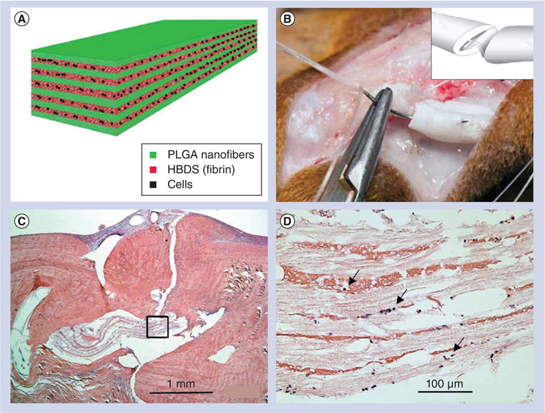
Figure 4. A layer-by-layer scaffold composed of poly(l-lactide-co-glycolide) nanofibers and a heparin/fibrin-based delivery system containing growth factor PDGF-BB along with adipose-derived mesenchymal stem cells for flexor tendon regeneration in dogs.

(A) The layer-by-layer structure of scaffolds. (B) The scaffold was grasped by a core suture and secured within the repair site. Inset shows that the scaffold was secured within the repair site. (C) Hematoxylin and eosin staining showing no obvious inflammatory response to the implantation of the scaffold 9 days postoperatively. (D) Magnified region in (C) showing only a small number of immune cells infiltrated the scaffold (arrows).
HBDS: Heparin/fibrin-based delivery system; PLGA: Poly(l-lactide-co-glycolide).
Reproduced with permission from [90].
