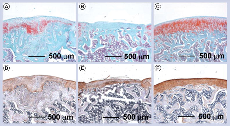
Figure 6. Electrospun poly(vinyl alcohol)-methacrylate/chondroitin sulfate nanofiber scaffolds for cartilage repair.

After implantation for 6 weeks in a rat osteochondral defect model, (A–C) safranin-O staining indicated that (A) the fiber implants promoted significant proteoglycan deposition compared with (B) the negative control (without treatment), while (C) native cartilage had the largest amount of proteoglycan deposition. (D–F) Immunohistochemical staining indicated that even (D) chondroitin sulfate fibers induced higher type II collagen production compared with (E) empty defects, but (F) native articular cartilage still contained significantly more type II collagen.
Reproduced with permission from [74].
