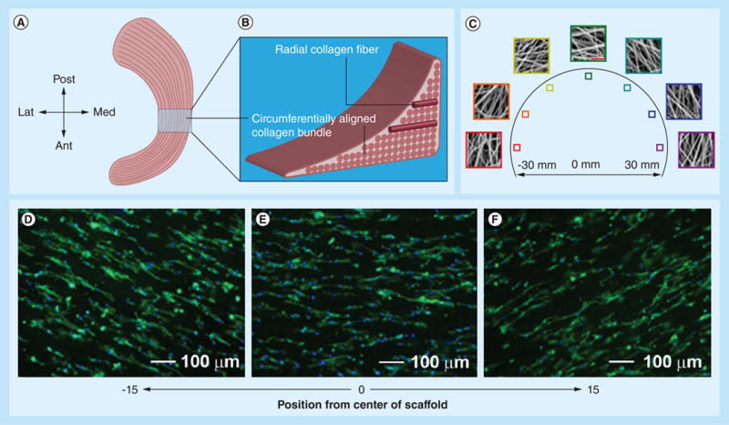
Figure 7. Fabrication of circumferentially aligned poly(ε-caprolactone) nanofiber scaffolds for knee meniscus tissue engineering.

(A) Anatomic macrostructure of meniscus. (B) A wedge-like cross-section displaying a simplified collagen fiber organization, with the majority of fiber bundles in the circumferential direction with occasional radial ‘tie’ fibers. (C) Scanning electron micrograph images showing different locations of samples for circumferentially aligned scaffolds (scale bar: 5 μm). (D–F) Fluorescent microscopy images of actin (green) and nuclei (blue) in juvenile bovine mesenchymal stem cells seeded on the different portions of circumferentially aligned scaffolds.
Ant: Anterior; Lat: Lateral; Med: Medial; Post: Posterior.
Reproduced with permission from [108].
