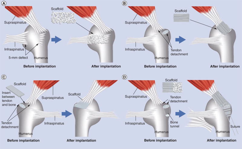
Figure 9. State-of-the-art nanofiber scaffolds for repairing rotator cuff injury in vivo.

(A) The scaffold (5-mm width and 5-mm length) composed of random poly(l-lactide-co-glycolide) (PLGA) nanofibers was used to bridge the gap between the infraspinatus tendon and humerus in a rabbit rotator cuff injury model. (B) The scaffold composed of aligned poly(ε-caprolactone) nanofiber scaffolds was implanted at the site between the supraspinatus tendon and humerus in a rat rotator cuff injury model. (C) The biphasic nanofiber scaffold composed of an aligned PLGA nanofiber layer and a PLGA–hydroxylapatite composite nanofiber layer was inserted between the bone and the detached tendon for integrative rotator cuff repair in a rat rotator cuff injury model. (D) The poly(ε-caprolactone) nanofiber scaffold with dual gradations in fiber organization and mineral content was used to repair rotator cuff injury in a rat shoulder model by inserting the mineral end into the bone tunnel and suturing the aligned end to the tendon.
