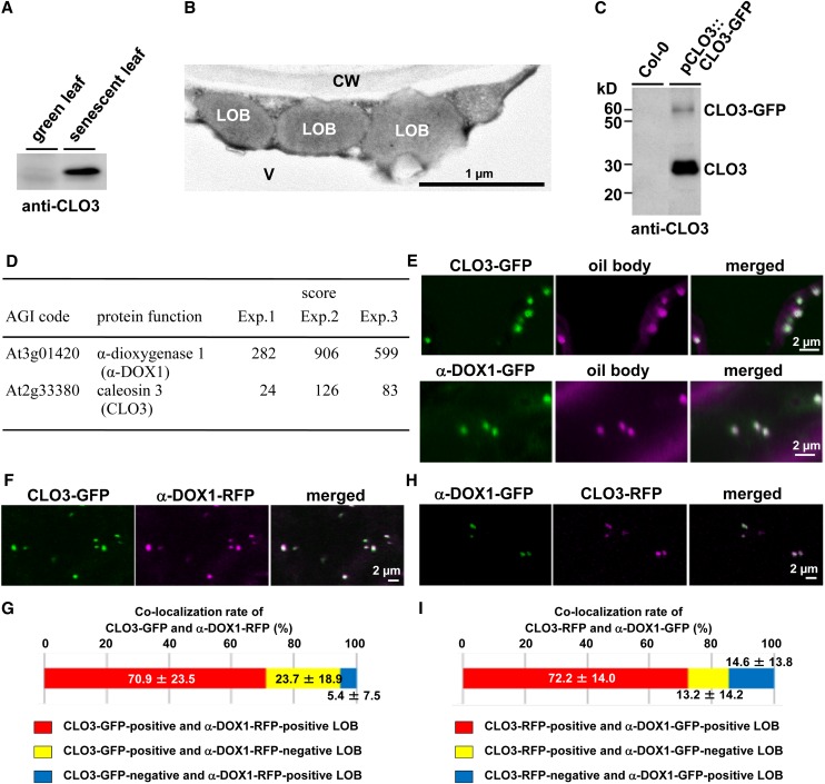
Figure 1.
Colocalization of CLO3 and α-DOX1 on the leaf oil bodies. A, Immunoblots showing the induction of CLO3 by senescence. B, Electron micrographs of senescent leaves showing spherical leaf oil bodies in cytoplasm. CW, Cell wall; LOB, leaf oil body; V, vacuole. C, Immunoblots showing that the pull-down CLO3-GFP-tagged oil bodies contained both CLO3-GFP and endogenous CLO3. D, LTQ-Orbitrap MS of CLO3-GFP-tagged oil bodies identified CLO3 and α-DOX1. Arabidopsis Genome Initiative (AGI) codes and annotations are from The Arabidopsis Information Resource database (http://www.arabidopsis.org). Scores were calculated using the Mascot program (Matrix Science). Raw data are given in Supplemental Data S1. E, Localization of CLO3 and α-DOX1 to oil bodies stained with Nile red. Either CLO3-GFP or α-DOX1-GFP was transiently expressed in N. benthamiana leaves. F, Colocalization of CLO3-GFP and α-DOX1-RFP by transient expression assay in N. benthamiana leaves. G, Quantitative analysis of the colocalization rate of CLO3-GFP and α-DOX1-RFP in seven fluorescence images. A total of 229 oil bodies were examined, most of which (yellow) were CLO3-GFP- and α-DOX1-RFP-positive oil bodies. Data represent average values ± sd. H, Colocalization of CLO3-RFP and α-DOX1-GFP by transient expression assay in N. benthamiana leaves. I, Quantitative analysis of the colocalization rate of CLO3-RFP and α-DOX1-GFP in nine fluorescence images. A total of 188 oil bodies were examined, most of which (yellow) were CLO3-RFP- and α-DOX1-GFP-positive oil bodies. Data represent average values ± sd.

