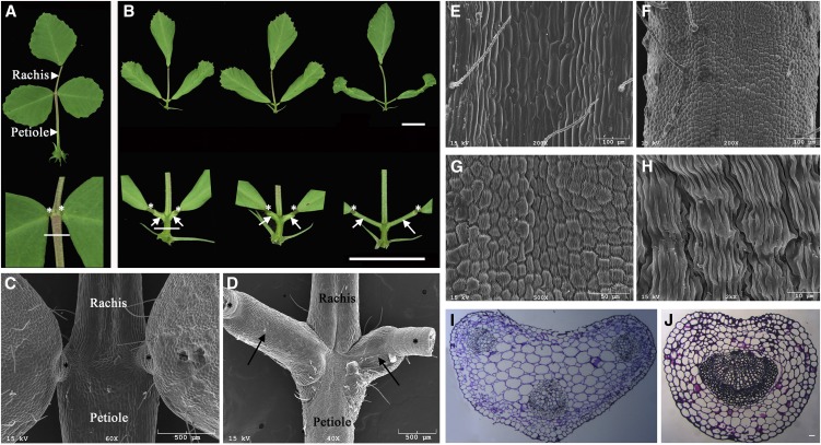
Figure 2.
Development of ectopic petiolules and altered petioles in mtphan plants. A and B, Representative 70-d-old wild-type (A) and mtphan mutant (B) compound leaves. Closeup images are shown at the bottom. Horizontal lines indicate the regions where SEM (E–H) and cross sections (I and J) were made. Bars = 1 cm. C and D, SEM images of wild-type (C) and mtphan mutant (D) compound leaves. Asterisks indicate pulvini, and arrows indicate ectopic petiolules. E to H, SEM images of petioles of 70-d-old wild-type (E) and mtphan mutant (F–H) plants. I and J, Cross sections of petioles of 70-d-old wild-type (I) and mtphan mutant (J) plants. Bar = 100 μm.

