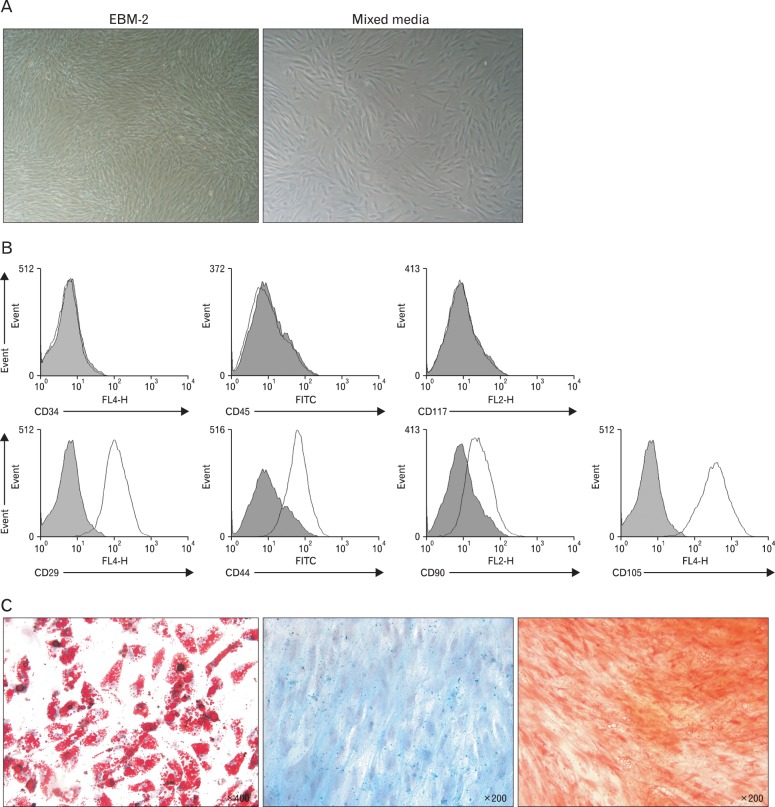
Fig. 1.
Establishment of human mesenchymal stem cells (MSCs) from adipose tissue. MSCs were isolated from human fat tissue. (A) On day 3 of culture, cells adhered to the bottom of the culture dish and appeared spindle-shaped. On day 3, cells cultured in EMG-2 media were nearly 90% confluent (left panel), while cells in mixed media were about 50% confluent (right panel). (B) The obtained cells were stained with antibodies against various surface markers, as indicated, and analyzed by flow cytometric analysis. Cell populations were negative for hematopoietic markers (CD34, CD45, and CD117), and positive for stem cell markers (CD29, CD44, CD90, and CD105). (C) Cells were induced to differentiate to adipocytes (left panel), chondrocytes (middle panel), or osteoblasts (right panel) using corresponding differentiation medium, fixed with 4% paraformaldehyde at an appropriate time point, and stained with Oil Red-O, Alcian blue, or Alizarin red S, respectively.

