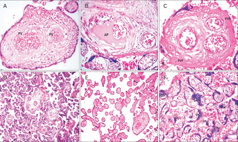
Fig. 1.
Histological sections of stem and terminal villi of preeclamptic placentas stained with hematoxylin and eosin. (A) A section of stem villus showing perivasculitis (PV) of fetal vessels. (B) A section of stem villus showing thrombosis and atheromatous plaque (AP) formation in fetal vessels. (C) A section of stem villus showing perivillous fibrin (PVF) deposition. (D) A placental section showing intervillous fibrin (IVF) deposition. (E) Avascular terminal villi of preeclampsia. (F) Placental villi showing the clusters or sprouts of syncytiotrophoblast, that is, syncytial knots (SK) (A-C, ×1,000; D-F, ×400).

