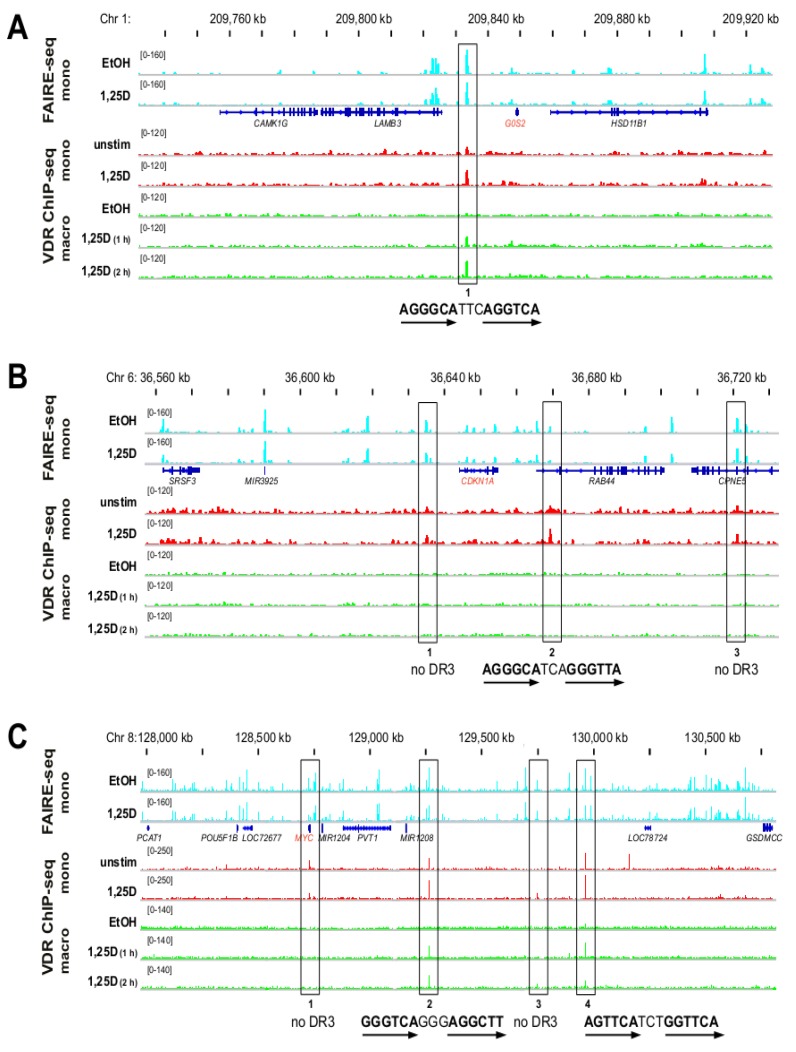
Figure 2.
1,25(OH)2D3-dependent chromatin opening and VDR association in monocyte- and macrophage-like cells. The IGV browser was used to display the VDR peaks of the chromosomal domains (see Supplementary Figure S3) of the genes G0S2. (A) CDKN1A (B) and MYC (C). The peak tracks show FAIRE-seq data obtained from monocyte-like cells (mono, undifferentiated THP-1 cells, treated for 100 min with EtOH or 1,25(OH)2D3 (1,25D), light blue [34]) in comparison to VDR ChIP-seq data from monocyte-like cells (undifferentiated THP-1 cells, without or with 1,25(OH)2D3 treatment for 40 min, red [4]) and from macrophage-like cells (macro, PMA-differentiated THP-1 cells, treated with EtOH or 1,25(OH)2D3 for 1 and 2 h, green). The gene structures are shown in blue. Investigated VDR peak regions are boxed. The sequences of the DR3-type VDR binding sites below the summits of the peaks are indicated (arrows indicate the hexameric nuclear receptor binding sites); some peaks carry no DR3-type sequence.

