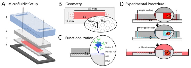
Figure 3.
Proposed microfluidic setup by Bichsel et al. [97] that allows cancer cells to be transferred from a capturing microchip device to a cell culture dish. (A) Microfluidic setup with transparent cover (1), cover with inlet and outlet tubing (2), microstructured PDMS on a glass slide (3) and metal support frame (4); (B) Detail of the microstructured PDMS geometry containing pillars of 100 µm in diameter; (C) PDMS functionalization principle; (D) Schematic of sample loading in the microfluidic device and magnification of a pillar-bound cell, followed by injection of the hydrogel precursor that solidifies and encapsulates the cells. Gel-encapsulated cells are finally transferred to cell culture conditions to be grown. Reproduced from [97] with permission from The Royal Society of Chemistry. For more details, the reader is referred to the original publication.

