Figure 4.
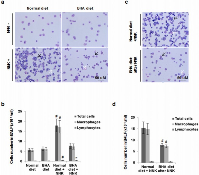
BHA blocks NNK-induced increase of macrophages in BALF. (a) A/J mice were maintained on normal or BHA diet for 2 weeks prior to NNK injection. BALF cells were prepared and stained with Wright and Giemsa after two weeks after NNK treatment. Representative images are shown; (b) Quantitative analysis of (a) is shown. The percentages of leukocyte types in BALF were determined by counting 400 leukocytes in a randomly selected portion of the slide under light microscopy; (c) A/J mice were maintained on normal or BHA diet 1 week after NNK treatment. Representative images of BALF cells collected 2 weeks after the last NNK dose. Represetitive Wright-Giemsa stainings are shown; (d) Quantitative analysis of (c) is shown. Black arrow: macrophage; White arrow: lymphocytes.

