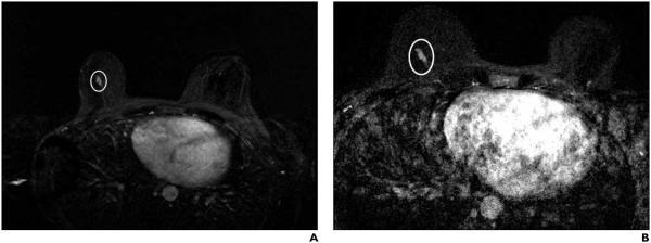
Fig. 2.

45-year-old woman who underwent bilateral breast screening MRI.
A, On bilateral dynamic contrast-enhanced breast MRI with subtraction, focal area of nonmasslike enhancement (oval) in lower central right breast was recommended for MRI-guided biopsy. MRI-guided biopsy was canceled. Patient underwent serial follow-up breast MRI.
B, Bilateral dynamic contrast-enhanced breast MRI with subtraction performed 2 years and 1 month after canceled MRI-guided biopsy found no interval change in morphologic features or size of focal area of nonmasslike enhancement (oval) in lower central right breast, after accounting for difference in FOV. Finding was considered benign and no additional follow-up breast MRI was recommended at that time.
