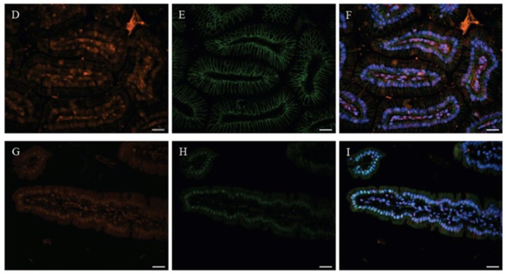
Figure 2.
Immuno-histochemical analyses in duodenal villi of mice. A to F, immunoreactive CaBP-9k appears in the cytoplasm of many populations of epithelial cells within the duodenal villi (A and D, red) at 100× magnification by immunofluorescence detection. To identify the co-localization between CaBP-9k and other proteins, the specific antibodies for chromogranin A (B, green) and E-cadhedrin (E, green) were co-incubated with anti-mouse CaBP-9k antibodies using corresponding Alexa-Fluor conjugated secondary antibodies. DAPI (blue) was used for nuclei staining. Based on DAPI signals, co-localization of CaBP-9k and each protein is evident by superimposition (C and F) of the green and red signals, respectively. HNF-4α (nuclear protein marker, G, Red) used in an attempt to identify the GR (H, green) expression in duodenal enterocytes, both images were merged and observed as blue (I). Scale bar = 20 μm.


