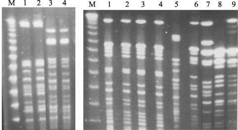
FIG. 3.
(Left panel) Gel picture showing two different digest patterns of MDR serovar Typhi from Kenya: PFGE pattern 1 (lanes 1 and 2) and PFGE pattern 2 (lanes 3 and 4) with SpeI restriction endonuclease. Lane M, 50-kb lambda molecular size ladder. (Right panel) Gel picture showing XbaI digest pattern I of MDR serovar Typhi from Kenya (lanes 1 to 3), a similar PFGE pattern of MDR serovar Typhi from South Africa (lane 4) but different from the digest patterns of MDR serovar Typhi from Hong Kong (lane 5), Pakistan (lane 6), and the fully sensitive serovar Typhi from 1988 to 1990 outbreaks in Kenya (lanes 7 to 9). Lane M, 50-kb lambda molecular size ladder.

