Figure 2.
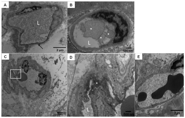
Mexico City dogs myocardial blood vessel pathology. (A) Scattered left and right ventricle capillaries exhibited endothelial cells with very dark cytoplasm, shrinkage, and partial detachment from the vessel lumen. This picture corresponds to a 4.6 years South MCMA animal facility male dog. “L” is marking the vessel lumen, magnification ×14,500; (B) South MCMA female dog with membranous luminal fragmented material in the vessel lumen (white *), magnification ×29,100; (C,D) correspond to a male 5 years old dog with numerous fine and ultrafine particulated material in the arteriolar wall (square), magnification ×7290; At ×29,100 (D), the conglomerates de PM are occupying the endothelial cytoplasmic space (white *); and (E) Normal left ventricle capillary in a 5 years old control dog, magnification ×14,500.

