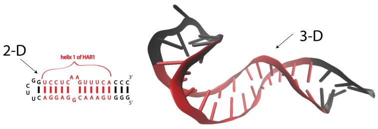
Figure 1.
Secondary (2-D) and tertiary (3-D) folds of helix 1 of human HAR1 RNA. Nucleotides, denoted in red, belonging to a native helical region of HAR1 RNA. Nucleotides, denoted in black, are added in order to stabilize the entire construct for NMR. The NMR structure is adapted from Ziegeler et al. [26].

