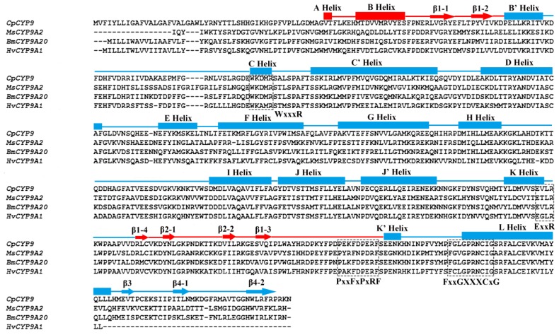
Figure 2.
Structure-based sequence alignments of Cydia pomonella CYP9A61, Manduca sexta CYP9A5, Heliothis virescens CYP9A1 and Bombyx mori CYP9A20 by ClustalW2. The predicted α-helices and β-sheets of CYP9A61 are marked on the top of sequences using boxes and arrows respectively. The β-domain is red, and α-domain is blue. The conserved regions of P450 enzymes including FxxGxxxCxG, PxxFxPxRF, ExxR, WxxxR are boxed using dashed lines.

